

Subscribe to RSS
DOI: 10.1055/s-0045-1814151
First X-Ray Facility in Asia: 125 Years of Excellence
Authors

Abstract
Medical radiology has a historical background in the discovery of X-rays (1895), by Wilhelm Conard Roentgen (1845–1923), who was awarded the first Nobel Prize in physics in 1901. Civilian X-ray facility was started in India in 1900 at the General Hospital, Madras (Chennai). Captain Thomas William Barnard was appointed as radiologist to the Government of Madras in 1920 to organize radiological service to the presidency. It was upgraded as Madras Government X-ray Institute (MGXI), in turn Barnard Institute of Radiology (BIR) in 1934. Radiotherapy for cancer treatment was started in 1924 and it was one of the four radium institutes in India (1934). Major V. Arthur Ponnaiah was the radium safety officer/first physicist who regulated radium storage, transport, and safety. The institute started its academic programs in radiology in 1936, first in Asia, and conducted research on age determination and forensic applications, first in the world. This X-ray facility has completed 125 years of public service and this article gives an overview of its origin and history at Madras (Chennai). Published literature, hospital records, souvenir articles, media information, and personal experiences are used to compile the present article.
Keywords
history of X-rays - Madras General Hospital - Captain Barnard - pioneer X-ray department in India - first X-ray image - Barnard Institute of RadiologyIntroduction
The General Hospital (GH) in Madras (Chennai) had the civilian X-ray facility in 1900, the first in South East Asia.[1] Captain Thomas William Barnard (TWB), United Kingdom, organized the radiological service for the Madras presidency ([Fig. 1]). The X-ray facility became the Madras Government X-ray Institute (MGXI, 1922), relocated and renamed as Barnard Institute of Radiology (BIR, 1934).[2] Radiotherapy for the treatment of cancer was started in 1924, followed by radium brachytherapy in 1934 and it was one of the four Radium institutes in British India. Medical and paramedical courses in radiology were started, first in Asia in 1936. Research on age determination and endemic fluorine poisoning based on X-ray investigation were undertaken. Radiographs of the institute were exhibited in London in 1921.[2] [3] The X-ray facility at Chennai (Madras) has completed 125 years of public service in 2025 (1900–2025). It is rare to find such an institute having radiodiagnosis, radiotherapy, radiological physics and nuclear medicine under one umbrella. Since there is a need to document the history of more than a century-year-old institution, an attempt is made to highlight it in an article for publication.
Origin and History (1900–1934)
Mahendralal Sircar (MLS; 1833–1904), a Bengali medical doctor and founder of the Indian Association of Cultivation of Science (IACS), had purchased an X-ray tube from Ducretet Company in Europe, within few months of its discovery. “The first experiment using X-rays in India was carried out by MLS on June 20, 1896 by taking the Radiograph of a hand using the procured Roentgen's apparatus and it was noted in his diary that he did not obtain a good picture in his first attempt, probably due to overexposure. He repeated his experiment and obtained a better X-ray photograph on June 23, 1896, which was the first successful radiograph in India.[1] [4]
Experiments with X-rays were continued at the IACS by Amrita Lal Sircar (son of Dr. Mahendralal Sircar) under the guidance of MLS with blocks of wood and books of different thickness, with sheets of iron, tin foil, and zinc foil, as has been recorded in the diary dated December 13, 1899. This was the beginning of X-ray research in India after the discovery of X-rays, and thus IACS has the distinction of introducing X-ray research in India.[1] Paradyot Kumar Tagore along with Father Lafont took an excellent Radiograph of the right hand of Earl of Elgin, the then Viceroy of India, wearing rings that was published in the Journal of Photographic Society of India in 1897.[1] C. V. Raman conducted research on X-ray scattering and diffraction by liquids, to study the structure of matter, thus paved the way for X-ray crystallography research in India (1921).[1] [4]
“As per available literature, the evidence of first clinical uses of X-rays in India, at least in an informal setting, was started by Acharya Jagadis Chandra Bose (1858–1937). After his Europe visit, he built an improved version of Roentgen's apparatus in the Presidency College, Calcutta (Kolkata, 1897–1898). Instead of connecting the Ruhmkorff's coil directly to the discharge tube as Roentgen did, he connected the Ruhmkorff's coil through a Tesla coil to the discharge tube, to get increased power. He mentioned his X-ray apparatus, in a letter written to Rabindranath Tagore in February 1898 (Sen, 1994, Roy, 2015) and requested him to visit the Presidency College to witness an examination of a patient with a broken back by using Roentgen kol (machine in Bengali).[1] He was also interested in the study of interaction of X-rays with various materials; created a barium platinocyanide screen to record the X-ray photographs of human hand, coins in a purse, etc., first in India (1898) and also founded an easily available potassium platinocyanide screen, substituting barium platinocyanide.[1]
Major Walter Calverley Beevor (Surgeon, 1858–1927) of the British army brought an X-ray apparatus from Britain and employed it in the Indian North West Frontier in the Army, which was the first radiograph taken in proximity to the battlefield by the British Army during the Tirah Campaign (1898) in northern India (Tirah Campaign was the Frontier war [1897–1898] in British India to regain the Khyber pass from the Afridi tribes).[5] Again used the X-ray facility in the Boer war (1899–1902, South Africa) along with Scots Guards in 1899 (https://www.victorian-cinema.net).[6]
Installation of the first X-ray machine in hospitals for public use is full of claims, counterclaims, and conflicting reports, so that it is very difficult to arrive at a definite conclusion.[1] [4] Dr. Kartick Bose (1873–1955), an eminent doctor, medical scholar, and industrialist, was the “first clinician in India to start clinical and X-ray laboratories.”[1] Madras Medical College claimed that the first X-ray outfit was obtained for the general hospital in the year 1900, the first in South East Asia. This might be true considering that it is a very old medical institution first started as a Government General Hospital on November 16, 1664 to treat sick soldiers of the East India Company ( www.mmc.ac.in ; accessed on June 17, 2021).[1]
Thus, GH, Madras, had the first X-ray apparatus installed in a hospital setup for civilian use in 1900, almost 5 years after the discovery of X-rays (www.mmc.ac.in/www.barnard,in).[2] [3] The X-ray facility was housed in two small rooms on the ground floor, western wing of then surgical department, GH (now demolished) for approximately 20 years. It was under the in-charge of assistant surgeon Rao Sahib M. Govindarajalu Naidu since 1909 (while senior surgeon and professor Lt. Col. John Maitland served at the GH) and it became a full pledged department in 1920.[2]
X-ray tubes employed at the GH were very primitive, coil unit operated from the mains and patients were exposed for several minutes. It had induction coils with various types of interrupters and several gadgets and devices to overcome the leakage of high-tension and inverse currents. In addition, the X-ray tube required optimal cooling by various means. They were single phase, self-rectified X-ray machines with air-cooled rectified valves, cones and cylinders, and their tables were either mechanically or manually operated with crude spot film devices. Multiple tubes of different penetrating power were used for different investigations. There was little protection against radiation and electrical hazards, and staffs were named as X-ray operators.
The first private X-ray facility at Madras (Chennai) was P. Rama Rao's X-ray institute (1910), located on Poonamallee High Road, Kilpauk. The X-ray unit of GH and that of P. Rama Rao managed various patients of the Madras Presidency, but the latter had major share of patients. The other government X-ray facility at Thanjavur was operated by a battery of 60 accumulators yielding approximately 110 volts. Batteries were recharged by means of an ancient dynamo and an ingeniously arranged equipment. The primary motive power was supplied by a Buffalo trotting round at one end of a long axle, while the other end is attached to a series of wheels, which when turned produced enough current to recharge the cells.[2]
In January 1920, TWB was appointed as a radiologist to the Government of Madras to organize the radiological service for the presidency (Madras presidency consisted of five southern states of India). He started his career as staff of The London Hospital, Whitechapel (1908), where patients were radiographed in their beds by means of portable trolley unit on which there was no protective shield attached to the X-ray tube. The X-ray tubes were 6 to 8 inch long, Gunderlach gas tubes were used, whose penetrating power was indicated by a Bauer Qualimeter. He operated the X-ray tubes at distance of approximately 10 feet to avoid radiation related injuries.
TWB came to India in August 1916 and installed an X-ray apparatus at the Cumbala War Hospital, Bombay, which was later used at Gallipoli Penisula (World War 1 military campaign, Turkey). He developed the method of cooling the film (to avoid film melting due to heat) so that it can withstand the X-ray heat; packing ice below and around the processing dishes. Based on the experience, he equipped the dark room with Frigidaire plant, which kept the solutions down to 60° to 65° F.
Later, he was transferred to the permanent Chief Military Hospital, at Colaba on back bay reclamation, Bombay (water body to the west of the original Mumbai's settlement, forming an arc between the peninsulas of Malabar Hill and Colaba). He worked with Captain Shorten, I.M.S., where he earthed the negative side of the tube and coil to prevent the damage of X-ray tubes from leakage current (Journal of the Roentgen Society/BJR, 1917). He left Bombay in January 1920 and went to London on four month leave, visited many X-ray departments, and met several eminent men who had the desire to set up professional standards for radiographers, which lead to the formation of “Society of Radiographers.”[2]
When TWB joined the Madras presidency in 1920, he felt the X-ray rooms were not only inadequate but also quit unfit for the workload, hence he convinced the Superintendent of the GH and got an annexe, originally designed for operation theatres. This was a large private bungalow in a fallow field, was taken for 2-year rent, which was purchased by the Government later, and became temporary building for the X-ray department. R. A. D. Graham and P. S. Babu Naidu were TWB's medical assistants along with Thothadri, a trainee.[2] [7] They were working with gas model X-ray tubes and electrical apparatus for Faradic and Galvanic massage and muscle testing. There were no lectures but the trainees were advised to observe the work of the staffs. Later, the department was upgraded as an institute and inaugurated on March 17, 1922 by the Governor of Madras, Lord and lady Wellington and named it as Madras Government X-ray Institute (MGXI) and a souvenir was released.[2] [7] [8] The institute was equipped with modern diagnostic, therapeutic and electrical treatment equipment. Her Excellency's hand was radiographed with the new apparatus. Colonel Thomas Henry Symons, the then GH superintendent, convinced the Government to buy a 200 kV X-ray unit for cancer therapy, worth of Rs. 20,000/-, this is the beginning of radiotherapy at Madras (Chennai) (1924). It was the first double coil unit installed in the East and housed in an annexe building specially designed and built for it.[2] [9] Though the Countess of Minto reported that Kolkata was the first radiotherapy center in India, opened on January 25, 1910 (as per the Foundation Stone), when radiotherapy treatment had started in Kolkata is not clear.[10] [11] In 1929, M. J. Santhanakrishna Pillai, Benjamin, Thothadri, and S. Sabapathy Pillai (sub-assistant surgeon) were the medical staffs of the X-ray institute and worked on rotation in different sections of the institute, to familiarize all types of work. TWB's post was made permanent in 1922, as “radiologist for the Madras Presidency” in turn the Director, Government X-ray Institute.[8]
X-ray departments were opened at Ootacamund (Ooty) and Madurai in 1922 and local mill owners donated the equipment and apparatus worth of Rs. 16,000 to Madurai.[2] R. A. D. Graham, assistant surgeon, was transferred to Madurai as in-charge of the X-ray department. In parallel, the Indian Radiological Association (IRA) was founded by Ajit Mohan Bose and Subodh Mitra at Calcutta (Kolkata) on April 21, 1931. It used to meet as section of Indian Medical Association (IMA) but its activities had declined due to World War-II.[12]
The second private X-ray facility at Chennai was the Premier Radiological Institute and Cancer Hospital by Kalpathi Rama Iyer Doraiswami who was conferred Padma Shri award for his pioneering work. It was initially located in Triplicane (1935) and later shifted to Dr. Radhakrishnan Salai (Edward Elliot's Road, 1948). It had both diagnostic and therapeutic facilities, including a superficial therapy, deep therapy X-ray (200 kV, 1952), followed by a Tele-Cobalt Machine (Theratron Junior, 1962) for cancer therapy. It also used radium tubes and needles until it is replaced by by Cs-137 manual after loading brachytherapy (1990).[3]
Madras General Hospital and Madras Medical College
The British established a medical facility for the benefit of personnel attached to the Madras Army-Military Hospital (MH) on November 16, 1664.[13] [14] [15] It was initially located at Fort St George, then relocated to Periamet, Poonamalle High Road (Narimedu, Hog's Hill) in 1772, named as General Hospital and opened to the public in 1899.[13] It was the first established western science-based hospital facility in India and became a teaching hospital of Madras Medical College. It conducted first medical forensic examination in India as early as 1693. It became a civil hospital in 1899, renamed as the Rajiv Gandhi Government General Hospital (RGGGH) in 2011 and completed 250 years in 2022.[13] During the expansion process, new eight storey buildings of 64,863 sq. feet (Tower Block 1 and 2, 2005) and 432,000 sq. feet (Tower Block-3, 2019) have been constructed (www.mmc.ac.in).
Madras Medical School was founded on February 13, 1835, by Rt. Hon. Sir. Frederick Adam, the Governor of Madras (Chennai). It became the Madras Medical College (MMC) on October 1, 1850 and celebrated its 175th anniversary in 2010. It is attached to the Government General Hospital, which has 2722 beds at present, one of the Asia's biggest treatment facilities. The MMC was affiliated to the University of Madras (1857–1988) and The Tamil Nadu Dr. M.G.R. Medical University (since 1988).[15] MMC offers five undergraduate degrees, 17 postgraduate diplomas, 25 postgraduate degrees, and 14 super-specialty degrees. Many institutions like the Barnard Institute of Radiology (BIR), Institute of Obstetrics and Gynecology, Institute of Child Health, Institute of Mental Health, Regional Institute of Ophthalmology (1819, first eye hospital in India and second oldest in the world), and Institute of Rehabilitation Medicine are attached to the MMC for teaching and training.
This college had the distinction of graduating one of the first four women doctors in the world, Mary Scharlieb (1878) who had higher studies at the Royal London School of Medicine, United Kingdom. She returned to India and started the Government Kasturba Gandhi Hospital for Women and Children (Royal Victoria Hospital for Caste and Gosha Women) at Madras (Chennai) and also established the Women's Medical Service in 1916 (medical graduate from MMC was eligible to register and practice in the United Kingdom and other colony countries). Muthulakshmi Reddy, who founded the Adyar Cancer Institute (WIA), was the first Indian woman doctor graduated from MMC (1912) and the first woman nominated to a Member Legislative Council (1916).[15] M. R. Gurusamy Mudaliyar, awardee of Chipperfield God Medal, passed general medicine (1920) and obtained special training in radiology at Dehradun. He was the Honorary Director of Indigenous System of Medicine (1937), first editor of the book, Materia of Medica (He was Honored with Father of Medicine), and first doctor to be awarded DSc by the University of Madras (1957). The first women Neurosurgeon in Asia, T. S. Kanaga was a graduate of MMC, who introduced stereotactic surgery and chronic electrode implantation in the brain. V. Shanta, the former Chairman of the Cancer Institute (WIA), Adyar and winner of 2005 Ramon Magsaysay award, was a graduate of MMC.[15]
MMC is known for several firsts in India; Institute of Radiology, (1934, TWB), Diabetic Clinic (1953, Sam G. P. Moses), Department of Urology (A. Venugopal), Cardiothoracic surgeon (A. Sadsivam), Institute of Neurology and Head Injury unit (B. Ramamurthy), Department of Surgical Gastroenterology and first MCh course (N. Rangabashyam), Department of Medical Oncology (S. Subramaniam, 1973), Department of Geriatric Medicine (V. S. Nataragan, 1978), and identification of first human immunodeficiency virus case in India. A. Lakshmanaswami Mudaliyar was the first Indian appointed principal of the college in 1939.[13] [15] Madras General Hospital and the Madras Medical College, currently spread over 47.5 acres of land with 52 operation theatres, 1,000 kVA generator, and 13,000 L of liquid oxygen facility and supporting approximately 12,000 outpatients per day.[15]
Barnard Institute of Radiology (1934–1969)
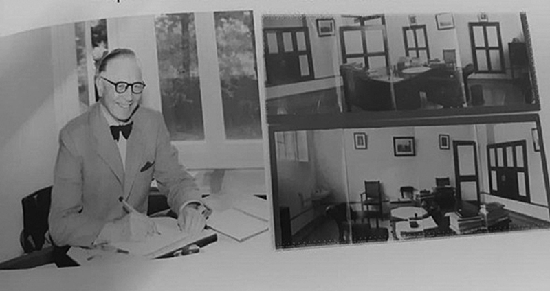
The Madras Government X-ray Institute was shifted to a new permanent building, which was constructed at a cost of Rs 3.29 lakhs. It was inaugurated on March 26, 1934, by Sir George Frederick Stanley, Governor of Madras, and a preliminary inaugural note on the institute was released ([Fig. 2A–C]).[2] The new institute was named as Barnard Institute of Radiology (BIR) by the Governor against the original proposal “The Madras Government Institute of Radiology (MGIR)” and TWB became its founding director. The British Journal of Radiology published an inaugural note of the institute as in the insert.[16]
“The Madras Government Institute of radiology, opened on March 26, 1934, by His excellency the Governor of Madras, provides the Presidency, and indeed the whole of India, with one of the most up-to-date radiological services in the world.
The institute, which forms the part of the scheme for re-building the Madras General Hospital and Medical College, is fortunate in having a Director Captain T.W.Barnard, well known for his pioneer work and long service in the cause of Indian radiology.
The well-equipped departments are staffed by fully-trained radiologists and assistants, and there are research laboratories, including a radon laboratory, for which it is hoped that the Government will allot 200,000 rupees worth of radium during the years 1935-36.
In 1932, a Radium Therapy and Research Committee was set up on the lines of that appointed by the Privy Council Medical Research Council in England.
It is hoped that the near future to have established a full course for Diploma in Medical Radiology, and to have created a Chair of Medical radiology in Madras University.
Arrangements are being made for the training of radiographers, with a certificate some- what similar to that offered by the Society of Radiographers”.

BIR had departments of radio-diagnosis, radiotherapy and electrology, radiological physics, and clinical photography with wide corridors, an elevator to transport patient, and a library of 500 books and 7 journals, best in the world. K. Manjunath Rai (K. M. Rai), lecturer in radiology, was in-charge of radiotherapy and electrology and T. K. Sundaram, a private radiologist, trained in Vienna, was in-charge for radio-diagnosis on contract basis.[7]
The institute was equipped with a 400 kV X-ray unit (Maximar, GE) for cancer therapy, first installed in the East.[2] [3] Dewan Bahadur M. R. Subbiah Chettiar donated a primary (GE, Victor XP4) and a secondary screening X-ray unit.[7] Additional units like convergent beam, pendulum therapy, and another type of moving therapy were added long before major centers in England installed such units.[3] The Medical & Lay press in the United Kingdom and United States described BIR as one of the best radiological services in the world (BJR, June 1934).[3] [7]
Radium Institute
BIR had 216 number of tube and needles of radium of strength 595.5 mg along with a radium storage safe and was one of the four radium institutes in India apart from Ranchi, Agra, and Lahore (Pakistan).[10] [17] Colonel Vaughan J. C. used radium in Ranchi in 1913, which was later shifted to Patna, attached to the Patna Medical College, the first in India. The Agra Radium Center was established in 1932 as Laxmi Panna Lal Radium Institute (Sarojini Naidu Medical College & Hospital), where P. K. Haldar was in-charge of radium, later.[17] Lahore Radium Institute (Pakistan) was founded in 1940 (King Edward Medical University).[18]
TWB got two water-colored sapphires from the jeweler Vummidi Bangaru, Madras (Chennai) on loan and irradiated them with radium gamma rays to enhance the color of stones (1937). After 24 hours, stones changed into a brilliant golden color and appeared to be topaz. Since the expected cornflower blue sapphire was not obtained, the trial was suspended.[2]
Once a 2-mg radium needle was missing during removal of an interstitial implant from a patient of carcinoma cheek. Physical search in the radium ward totally failed, but the GM counter survey identified the needle at 10 feet height above the patient. A crow could have picked the needle and dropped on the fan blade. Similarly, the Government Women and Children Hospital, Egmore, Madras (Chennai), missed 20 mg of radium tube used in the intracavitary application of the cervix. The physicist from BIR was deputed to investigate, finally the radium tube was recovered from a garbage dump outside the hospital after 3 days—says C. Rethibnasapathy.
Education and Training
Post-Diploma in Medical Radiology (1-year DMR) was started in 1936 under the Director of Medical Education, first time in Asia. K. A. Kannan, Y. Siddique, A. N. K. Menon, and Arthur Daniel were the first graduates of the course, who were later absorbed as assistant surgeons for the Madras Presidency. I. S. Sodhi of Chandigarh, S. V. Tungar of Nagpur, T. Jayanarasimhulu, and T. R. Seshagiri Rao of Andhra Pradesh were also trained in the institute.[8] Post-Diploma in Medical Radiology (2-year DMR) was started in 1956 under the University of Madras, first in Asia. Dark-Room Assistant (Six-month DRA) and Certified Radiological Assistant (One-year CRA) training were started in 1936, first in Asia. TWB was appointed as the first president of Board of Examiners for the diplomas.[2] Chief radiographer Tholasingam was in-charge of maintenance of all diagnostic, therapeutic, and physiotherapy instruments including other hospitals in the city and state. Ms. Beulla was the radiographer who later qualified MBBS and FFR (Lond.) and became Reader in diagnostic radiology, who later shifted to Christian Medical College, Vellore.[7] [8]
Research Schemes
The institute performed two research schemes on (1) determination of the age of a patient by a series of 500 radiography examinations of the epiphysis, and (2) investigation on endemic fluorine poisoning by large number of X-rays. TWB cited some cases of investigative X-ray radiography: (i) radiography showing a gold chain lying in the stomach of a thief, which was snatched from a small girl during a temple festival, (2) radiographs showing precious stones, which were found secreted in small cavities inside the cheeks of a women, who was suspected a member of criminal band, (3) testing and certification of a 500-year-old large diamond by X-rays that belonged to the Indian Prince in the North, (4) radiograph showing outlines of small nails and mercury from a fakir's stomach, and (5) X-ray examination of sprained ankle of a potential smuggler, which revealed the jewel of a lady passenger on a ship—the smuggler's heel of the shoe was hallowed and filled up with jewels[2] ([Fig. 3]).

In June 1921, 24 radiographs from the X-ray Institute, Madras were exhibited at the Roentgen Society, and the best three were shown again at the Royal Photographic Society of Great Britain, London in September and October. Over the period, approximately 20 X-ray facilities were opened in the Madras Presidency and were supervised by TWB, which is more than three times the number required for the territory of Britain with a population of 50 million.[2]
K. Manjunath Rai
K. Manjunath Rai (K. M. Rai) was the first Indian director who succeeded TWB in 1941 who started his career as assistant professor of operative surgery, went to the United Kingdom and qualified DMR (Lond.) and FRCS (Edin.), and later worked in the Government Women and Children Hospital, Egmore, before joining as lecturer in BIR ([Fig. 4A]). K. M. Rai along with P. K. Haldar (Agra), J. P. Sinha (Patna), and M. L. Aggarwal (Amritsar) had undergone special training in the use of radium and X-ray therapy in the United Kingdom (Great Britain).[19] K. M. Rai was instrumental for the installation of two short wave units (M/s. Powells Ltd, Bombay and M/s.Philips,1957).

Before 1948, radiologists has no independent status, all cancer patients were admitted under the control medical and surgical wards and referred to radiology for treatments based on their instruction, including deep therapy X-ray fractions and number of radium needles, etc. K. M. Rai got a separate cancer unit with 100 patients capacity including a radium ward attached with an operation theatre and anesthetist, which was a milestone in the history of BIR and India ([Fig. 4B]). S. Krishnamurthy (Cancer Institute (WIA), Adyar) was in-charge of cancer OP on honorary capacity and T. K. Rithuparnan DMR, DMRT (Lond.), was a senior lecturer in radiology and radiotherapy (1948). The Madras Cancer Association donated a cancer pavilion from the provincial welfare fund and was opened during the occasion of the bi-centenary celebrations of the GH in 1953 ([Fig. 4C]). K. M. Rai started weekly cancer conferences consisting physicians, surgeons, radiotherapists, and pathologists. He also started cancer outpatient department and distributed cancer awareness pamphlets in four languages. He was responsible for the upgradation of BIR as all-India postgraduate training and research center (1958).
K. M. Rai was a founder member of the Academy of Medical Sciences, New Delhi.[7] P. Rama Rao, K. M. Rai, and Santhana Krishnan Pillai have revived the IRA and conducted the first Annual Congress of Radiology in 1946 at Madras (Chennai), under the presidentship of M. D. Joshi. P. Rama Rao became the secretary of IRA (1946) and founder-editor of the Indian Journal of Radiology along with K. M. Rai (1947).[12] Barnard Institute celebrated its silver jubilee in 1959 (1934–1959) under K. M. Rai (other members were Arthur Daniel, A. N. K. Menon, T. K. Rithuparnan, P. S. Babu Naidu, R. Tholasingam [chief radiographer], and Smt. J. Halibaran [staff nurse]). TWB wrote an article in the silver jubilee souvenir “X-rays-Personnel Collections,” which is a current source of information to all.[2] After retirement, K. M. Rai established the Rai Medical Center, Teynampet, Chennai in 1962 and practiced radiotherapy with radium tubes and tele-cobalt therapy.
Y. Siddique
Y. Siddique succeeded K. M. Rai as director and head of radiotherapy (1961–1968) whereas M. G. Varadarajan was in-charge of radiodiagnosis ([Fig. 5A]). Y. Siddique and A. D. Singh (Patna) had undergone training in radiation therapy in England and were awarded the Radiology Fellowship (FFR), London. Y. Siddique opened the outpatient X-ray block and commissioned a 300-kV superficial therapy machine (Stablipan, M/Siemens, 1964) and a cobalt therapy unit, Gammatron-2 (M/s. Siemens, 1967).

Post-Diploma in Medical Radio-diagnosis (DMRD, 1962), and Radiotherapy (DMRT, 1962) and MD (Radio-diagnosis and Radiotherapy combined, 1963) were started under the University of Madras, first in Asia. Separate MD courses in radio-diagnosis and radiotherapy were started in 1967 under the same university. Radiological physics was a separate theory paper with practicals followed by the university examination, first in India.[8]
Y. Siddique was instrumental in starting a nuclear medicine department at the institute in 1962. C. Rathinasabapathy, the second physicist, was trained at the Radiation Medicine Centre (RMC), Bhabha Atomic Research Center (BARC), Bombay, in 1965. K. R. Rajagopalan, radiologist, had his clinical nuclear medicine training in the United Kingdom in 1966.[2] [3] Y. Siddique died on February 29, 1968 while in service, and he was succeeded by A. N. K. Menon.[17]
Major V. Arthur Ponnaiah
Major V. Arthur Ponnaiah (1934), was the first physicist, in-charge of radium warehouse, physics laboratory, and mold room ([Fig. 5B]). He established physics museum and control of various protective measures needed for the institute against X-rays and radium. He used to see that the rules regarding the custody, issue, use, and receipt were strictly complied with, all linked with radium and bring to the notice of the Dean, any violation of the rules laid down for the protection of the staff and any improvement, if need be, in this regard. He was also officer-in-charge of stores and supervised the store section from the technical point of view and regulate the receipt and issue of stores.[20] [21] Thus, Major Ponnaiah was the first Medical Physicist in India.
Barnard Institute of Radiology and Cancer/Oncology (1970–2000)
Arcot Gajaraj
BIR was renamed as Barnard Institute of Radiology and Cancer (BIR&C) in 1969, to justify its role in both radiodiagnosis and radiotherapy and Arcot Gajaraj became first Director (1970–74 and 1976–79) ([Fig. 6]). Arcot Gajaraj had his training at London (DMR, FRCR) and introduced percutaneous lumbar aortography and venous angiocardiography. He served as president of Society of Nuclear Medicine, India (SNMI, 1977) and Indian Radiological & Imaging Association (IRIA, 1985) and as chairman of the Indian College of Radiology Imaging (ICRI, 1981)

A cesium teletherapy (M/s.Siemens, 30 cm SSD, 1972), ultra-shortwave diathermy (M/s. Siemens, 1972), double-headed Janus tele-cobalt teletherapy (M/s. IGE, 60 SSD, 1978), 100 kV superficial therapy (M/s. Phillips, RT-100, 1978), and a contact X-ray therapy (M/s. Siemens, Dermopan, 1979) were installed during his tenure. Arcot Gajaraj served as a member in the Radium Replacement Committee formed by the Atomic Energy Regulatory Board (AERB), Government of India.
Further Developments
First treatment planning system (Rad Plan, TSG, 1987) and BARC manual after loading Cs-137 kit for gynecological treatment (1989) and iridium-192 manual radioactive wire interstitial implant (1990) were put in to patient use. All the radium (595.5 mg) were surrendered to Indira Gandhi Center for Atomic Research (IGCAR), as per the directive of AERB along with radiums of Government Women and Children Hospital (780 mg) and Government Stanley Hospital, Chennai (1988).[17]
BIR&C was renamed as Barnard Institute of Radiology and Oncology (BIR&O) in 1991, thanks to M. Jagadeesan, the professor of radiotherapy. The Tamil Nadu M. G. R. Medical University approved the physics faculties to do part time PhD in radiology. A tele-cobalt unit (Phoenix, 1994) and a remote after loader for brachytherapy (Microselectron, Ir-192, 1997) were installed by the initiative of Prof M. Jagadeesan. BIR&O had its 58th year Annual Day celebration on June 26–27, 1992 (M. Panchatcharam) and Diamond Jubilee on March 26, 1994 (M. Jagadeesan).
Radiology Specialist
Arther Daniel, P. S. Babu Naidu, and Chinnaswamy were the first angiographers, and performed invasive procedures. First percutaneous nephrostomy, abscess drainage, and embolization of hand Arteriovenous malformation (AVM) and percutaneous transhepatic biliary drainage (PTBD) were done in 1978 by J. R. Daniel. M. G. Varadharajan was trained in neuro-radiology in Queen Square, London, by Prof. J. W. Bull and C. R. Jayakumar was in cardiovascular and vascular radiology under Robert E. Steiner and M. J. Rapheal at Hammersmith Hospital, London (1972–74). Arcot Gajaraj and A. S. Aswathaman were the forerunners in vascular and experimental angiography in animal models. Pediatric radiology was started by Vasanthkumar Shetty who had training under Prof. John Caffey.[8] P. C. Rajaram, cardiac radiologist, did pioneering work in lymphography, congenital anomalies of the heart, and special views for valves, etc., and obtained PhD and DSc in radiology from the University of Madras.
Barnard Institute of Radiology and Oncology (2000–2025)
J. R. Daniel and T. S. Swaminathan have initiated the BIR Worldwide Alumni Association on November 22, 2002, which was inaugurated by the Honorable Health Minister S. Semmalai, Government of Tamil Nadu. T. S. Swaminathan has initiated the installation of first 64-slice computed tomography (CT), and started the Barnard Institute of Radiology Alumni Advanced Continuing Education (BRACE) and Post-graduate-Primer course as annual events. Diagnostic radiology department was extended to the first floor of the newly constructed Tower block-I on July 1, 2005.[15]
Two-year Diploma in Radiology and Imaging Technology (DRIT) and Radiotherapy Technology (DRTT, 2009) were started under the Directorate of Medical Education (DME). BSc Radiography and Imaging Technology (RIT, 2012) and Radiation Therapy Technology (RTT, 2012) of 3-year duration plus 1-year internship courses were started under The Tamil Nadu M. G. R. Medical University.
Post Platinum Jubilee (PPJ) was celebrated on March 26, 2010 (M. Prabhakaran). Barnard's daughter, Mrs. Barbara King and grand-grand daughter, Ms. Rachel, United Kingdom, have participated in the PPJ celebration. They returned the silver souvenir (token gift), presented to TWB in 1940 during his retirement, which was engraved with all the employee's signature. Mrs. Barbara King wanted to see her father's garden home (Chipstead) at Nungambakkam High Road, Chennai, where she spent several years of her childhood. Hotel Taj Coromandal has come up on their garden house now (S. Muthaiah, Hindu articles 4, 2010).[23]
Presently, the department of radiodiagnosis is equipped with a bone mineral densitometer (BMD), a digital subtraction angiography machine (DSA), and 2 mammography, 19 radiography, 7 CT scanners, 8 ultra sonograms, 3 Doppler units, and 2 magnetic resonance imaging (MRI) machines. About 100,920 plain X-rays, 2,337 special procedures including angiograms, 105,421 CT scans including interventions, 91,759 ultra-sonograms including Doppler and guided biopsies, and 18,509 MRI scans are performed per year. Started with four employs in 1920, became 80 in 1940 and have 135 employees and 250 students at present, the only institute having a separate building as early as 1934 in the country.
P. Mahadevan Pillai, Santhana Krishna Pillai, Arthur Daniel, P. S. Babu Naidu, A. N. K. Menon, M. G. Varadarajan, A. S. Aswathaman, Arcot Gajaraj, Karkala Subbarao, K. R. Rajagopal, M. V. K. Shetty, Leela Meenakshi, G. Srinivasan, K. R. Rajagopal, P. C. Rajaram, C. R. Jayakumar, M. Sushilaraj, G. Munusamy, V. P. Narayanan, M. Jagadesan, I. Kandaswamy, D. R. Subramaniyam, J. R. Daniel, T. S. Swaminathan, and K. Thayalan are the notable alumni of the institute.
Departments of Radiation Oncology
K. M. Rai laid solid foundation for an organized radiotherapy department with X-ray therapy and radium after TWB. The successive heads of radiotherapy are Y. Siddique, K. Radhakrishnan, G. Srinivasan, Kamalabai Sampoornam, M. Jagadeesan, G. Narayanan, N. Thoppa Goundan, M. Balu David, N. Mohan Ram, S. Shanmugakumar, and T. N. Vijayshree.
The department is presently equipped with a High dose rate (HDR) brachytherapy unit (BeBig, Multi source, 2015), a dual-energy linear accelerator (Varian, True beam STX, 2019), CT simulator (Canon, 2019), and a tele-cobalt (Equinox, Team Best Asia LtD., 2024) including 15 beds for cancer intensive care unit. About 1,500 patients are treated per year including brachytherapy with state-of-the-art equipment, comparable to any corporate facility. It celebrated its centenary celebration (1924–2024) on September 13, 2024, during the inauguration of the fourth successive tele-cobalt unit (Equinox), in the presence of the Honorable Heath Minister Ma. Subramanian, Government of Tamil Nadu. A centenary celebration symposium (Master Class-II) was also conducted on December 7, 2024.
Tamil Nadu Medical Service Corporation (TNMSC) installed 11 linear accelerators, 15 tele-cobalt therapy, 7 HDR brachytherapy, and 9 CT simulator machines across the state, as part of their operate and maintain-therapeutic services in 2019. To strengthen the physics and radiation safety support, it recruited 34 medical physicists and Radiological safety officer (RSO)s for various government hospitals in the state including BIR&O.
Department of Radiological Physics
Department of radiological physics was established in 1934 and continues to offer services to both radiodiagnosis, radiotherapy, and nuclear medicine in terms of clinical, teaching, regulation, safety and research. Major V. Arthur Ponnaiah (1934–41) was the first physicist and he was assisted by a second physicist. The successive heads are V. Krishnamurthy (1947–56), T. M. B. Nedungadi (1962–79), C. Rethinasabapathy (1979–91), A. Thanumalayan (1992–95), K. Thayalan (1997–2012), K. Muthuvelu (2013–19), and P. Kaliyappan (2020–23) and Arumugam Kopperundthevi (2024).[21]
First physicist performed periodical physical verification of stores by surprise checks and supervised the preparation of developer and fixer solutions once a week. The second physicist used to calibrate of X-ray therapy apparatuses and support the radiotherapists in the calculation of tumor dose, etc. Physicists were also a general supervisory in charge of all the electromedical X-ray apparatus in the institute and their proper maintenance and gave help when needed by the chief radiographer in any breakdowns or other repairs. Wire man on duty assisted the physicists in the maintenance of transformer rooms and high-tension cables. A dairy was maintained for the inspection of the director.[8] The first and second physicists designation has been changed into professor and assistant professor of radiological physics in 1979. The present responsibilities include calibration of radiation generating equipment, cancer treatment planning, regulatory affairs related to AERB, radiological safety, and teaching.
Department of Nuclear Medicine
Department of Nuclear Medicine was started in 1962, headed by K. R. Rajagopalan, the radiologist.[20] [22] The successors are R. Natarajan, N. Ramadas, K. Vanitha, and E. Prabhu who were trained at the Radiation Medicine Center (RMC), Mumbai, with Diploma in Radiation Medicine (DRM). RIA, biochemistry laboratory, thyroid uptake probe, and high-dose iodine therapy with two-bed services were offered to patients, unfortunately no nuclear medicine service is available at present, though positron emission tomography/Computed tomography (PET/CT) facility is offered under public-private partnership (PPT), which carry out 6,000 scans per year.
T. W. Barnard, a Great Contributor in Radiology
After retirement in India, TWB returned to England (1942) and took in-charge of Sheffield Royal Infirmary as secretary of the Sheffield National Center for Radiotherapy. He worked with upcoming technologies like mega voltage therapy, isotope facilities, and founded a new radiotherapy hospital in 1948. He again retired in 1964 and held negotiations with Massachusetts Institute of Technology (MIT) to build a 2-MV Van de Graaff generator at Sheffield. He passed away in 1978 at the age of 93 after five decades of X-ray service both in India and the United Kingdom.
TWB once quoted “Although I used a naked X-ray tubes with no protective shield, I escaped series injury apart from damaged finger nails, as I took precautions ignored by my seniors, the most important being to keep a safe distance from the X-ray tube when it was in action: I attribute the fact that I am alive today being due to my use of a length of insulated flex by means of which I switched the tube on my ward apparatus ON and OFF from a distance of about 10 feet” (Maddy's Ramblings, February 9, 2019 [last accessed on September 12, 2025]).
“Captain Barnard belonged to the category of great men who had a modest beginning, but by dint of hard work, perseverance and foresight brought laurels not only on themselves but also made available contributions to the welfare of mankind. He was not a product of any medical school and began his life as an ordinary X-ray operator hardly 10 years after the very discovery of X-rays” – Obituary note by Arcot Gajaraj.
“There was nothing like it in India or in South Asia, and not much like it in the USA or Great Britain at that time. It (BIR) was a two storey building around a courtyard where playing fountains formed part of the air-conditioning plant of the X-ray diagnostic department. Its therapy unit housed a 400 kV apparatus, three 200 kV units, two superficial and two contact sets. It had a Radium department and a clinical photography unit. With its protected walls of locally made brick loaded with barium it had a layout so far in advance of its time that for a generation it remained the outstanding radiological center of South East Asia. The equipment has been kept up-to-date by adding new units such as Convergent beam, Pendulam and another type of moving beam therapy apparatus-long before important centers in England received such units” – Obituary note by H. Miller.
TWB was a treasurer and deputy chief commissioner of the Madras Boy Scouts Association, keen collector of art subjects, interested in gemology, and the identification of gem stones. He also helped four poor student girls (O. J. Annie, Marry, Catherine, and Elizabeth) of a small village near Trichur in Kerala, toward their food, books, and clothes based on their letter, August 8, 1923.
Conclusion
Civilian X-ray facility was started in Madras (Chennai) in 1900 and became an institute of radiology, first in Asia, which had all radiological sciences under one roof. It started radiotherapy for cancer in 1924 and one of the four radium institutes in India. BIR&O is decorated with TWB, K. M. Rai, Y. Siddique, Arcot Gajaraj, M. Jagadeesan, D. R. Daniel, and T. S. Swaminathan who made great strides in radiology. TWB's contribution is unique since he was not a doctor licensed with medical degree, but was virtually considered one by dint of his meritorious service and the knowledge he possessed about his own field. He often said: “even with poor equipment, you can get good results by dint of hard work.”
There is a major concern to recommission the nonfunctional nuclear medicine department, to bring back to its original status, along with latest molecular imaging facility. Newer teaching courses like DM in Neuroradiology and MSc in Medical Physics courses are overdue. Though a large volume of patients are handled at free of cost, the research and publication number has been not proportionate, due to increasing patient service, lack of funds, and commitment from the faculty, except few individuals. Long-term vision, policy making, and new research projects is the need of the hour.
The Barnard Institute of Radiology and Oncology (BIR&O), the forerunner institute, has been a pride during British and independent India, which has been in public health service over 125 years. This article is a standing tribute to TWB, the X-ray man of Madras who developed the radiological sciences on par with world standard and initiated many first in East Asia. There is a need to preserve the BIR&O's heritage by providing equipment and infrastructure and upgrade it a Center of Excellence of National Importance in Radiological Sciences.
Conflict of Interest
None declared.
Acknowledgments
The authors would like to acknowledge Dr. S. Kalpana, Professor & Director, Dr. T. N. Vijayashree, Professor & Head, Department of Radiation Oncology, and Mrs. Arumugam Kopperunthdevi, Professor of Radiological Physics, Barnard Institute of Radiology & Oncology, Rajiv Gandhi Government General Hospital & Madras Medical College, Chennai, for their contribution and support towards this article preparation.
-
References
- 1 Roy SC. History of X-ray research in Colonial India. Indian J Hist Sci 2018; 53 (04) T123-T131
- 2 Barnard CTW. X-rays-personal recollections. J Med Phys 1995; 20 (03) 5-9
- 3 Hemanth Raj E. How Cancer Care Developed in India in 75 Years of Independence-Tamil Nadu scenario. Indian J Surg Oncol 2022; 13 (Suppl. 01) S36-S41
- 4 Suprakash C. Roy. Discovery of X-rays- Its impact in India and history of X-ray research in colonial India. Quantum Beam Sci 2022; 6 (02) 16
- 5 Parthasarathy KS, Predeepkumar KS. . Evolution of Health and Safety Program 1987; BARC: 102–119. Accessed December 25, 2024 at: https://www.barc.gov.in
- 6 Vincent J. Cirillo, The Spanish-American War and Military Radiology. Am J Radiol 2000 174. (05)
- 7 Thayalan K, Amarnath C. Barnard Institute of Radiology and Oncology. Chennai: Post-Platinum Jubilee Celebrations-Souvenir; 2010: 28-33
- 8 Daniel JR. Barnard Institute of Radiology-a centre of excellence. Barnard Institute of Radiology World-wide Alumni Association. Chennai: Souvenir; 2002
- 9 Rethinasabapathy C. Memories of unforgettable saga. International Conference on Medical Physics and Radiation safety (AMPICON), Chennai: Souvenir; November 10–12, 2005:39–40
- 10 Munshi A, Ganesh T, Mohanti BK. Radiotherapy in India: history, current scenario and proposed solutions. Indian J Cancer 2019; 56 (04) 359-363
- 11 Banergee S. Setty UM, Shrivasta SK. Brachytherapy India-along road ahead. J Contemp Brachytherapy 2014; 6 (03) 331-335
- 12 Indian Radiological and Imaging Association. Accessed December 25, 2024 at: https://iria.org.in
- 13 Raman R, Raman A. On the quarter-millennial anniversary of the Madras General Hospital. Natl Med J India 2022; 35 (01) 47-51
- 14 Dorairajan N, Pandyaraj M, Muralidharan. et al. An ode to my alma mater - Madras Medical College. Indian J Surg 2007; 69 (04) 163-168
- 15 Gopal M, Balasubramanian D, Kanagarajah P, Anirudhan A, Murugan P. Madras Medical College: 175 years of medical heritage. Natl Med J India 2010; 23 (02) 117-120
- 16 The Madras Government Institute of Radiology. Br J Radiol 1934; 78 (07) 343
- 17 Thayalan K. Evolution of Radiotherapy in Tamil Nadu. 37th Annual Conference of Association of Radiation Oncologists of India (TN & P Chapter), Chennai: Souvenir; September 24–25, 2022
- 18 Mohsin Fareed M, Jamshed A, Butt S, Shaukat F, Nguyen PL, Ngwa WF. Evolution of radiation oncology in Pakistan. Int J Radiat Oncol Biol Phys 2019; 105 (01) 11-16
- 19 Gupta BD. History of radiotherapy and infrastructure. Int J Radiat Oncol Biol Phys 1996; 36 (04) 945-947
- 20 Daniel JR. Barnard Institute: Yesterday-Today and Tomorrow- Barnard Institute of Radiology World-wide Alumni Association. Chennai: Souvenir; 2002
- 21 Thayalan K. Department of Radiological Physics, Government General Hospital. Barnard Institute of Radiology World-wide Alumni Association. Chennai: Souvenir; 2002
- 22 Vanitha K. Department of Nuclear Medicine, Barnard Institute of Radiology World-wide Alumni Association. Chennai: Souvenir; 2002
- 23 Muthaiah S. . Madras Miscellany, The Hindu, Chennai; April 04, 2010. Accessed May 25, 2024
Address for correspondence
Publication History
Article published online:
20 January 2026
© 2026. Indographics. This is an open access article published by Thieme under the terms of the Creative Commons Attribution-NonDerivative-NonCommercial License, permitting copying and reproduction so long as the original work is given appropriate credit. Contents may not be used for commercial purposes, or adapted, remixed, transformed or built upon. (https://creativecommons.org/licenses/by-nc-nd/4.0/)
Thieme Medical and Scientific Publishers Pvt. Ltd.
A-12, 2nd Floor, Sector 2, Noida-201301 UP, India
-
References
- 1 Roy SC. History of X-ray research in Colonial India. Indian J Hist Sci 2018; 53 (04) T123-T131
- 2 Barnard CTW. X-rays-personal recollections. J Med Phys 1995; 20 (03) 5-9
- 3 Hemanth Raj E. How Cancer Care Developed in India in 75 Years of Independence-Tamil Nadu scenario. Indian J Surg Oncol 2022; 13 (Suppl. 01) S36-S41
- 4 Suprakash C. Roy. Discovery of X-rays- Its impact in India and history of X-ray research in colonial India. Quantum Beam Sci 2022; 6 (02) 16
- 5 Parthasarathy KS, Predeepkumar KS. . Evolution of Health and Safety Program 1987; BARC: 102–119. Accessed December 25, 2024 at: https://www.barc.gov.in
- 6 Vincent J. Cirillo, The Spanish-American War and Military Radiology. Am J Radiol 2000 174. (05)
- 7 Thayalan K, Amarnath C. Barnard Institute of Radiology and Oncology. Chennai: Post-Platinum Jubilee Celebrations-Souvenir; 2010: 28-33
- 8 Daniel JR. Barnard Institute of Radiology-a centre of excellence. Barnard Institute of Radiology World-wide Alumni Association. Chennai: Souvenir; 2002
- 9 Rethinasabapathy C. Memories of unforgettable saga. International Conference on Medical Physics and Radiation safety (AMPICON), Chennai: Souvenir; November 10–12, 2005:39–40
- 10 Munshi A, Ganesh T, Mohanti BK. Radiotherapy in India: history, current scenario and proposed solutions. Indian J Cancer 2019; 56 (04) 359-363
- 11 Banergee S. Setty UM, Shrivasta SK. Brachytherapy India-along road ahead. J Contemp Brachytherapy 2014; 6 (03) 331-335
- 12 Indian Radiological and Imaging Association. Accessed December 25, 2024 at: https://iria.org.in
- 13 Raman R, Raman A. On the quarter-millennial anniversary of the Madras General Hospital. Natl Med J India 2022; 35 (01) 47-51
- 14 Dorairajan N, Pandyaraj M, Muralidharan. et al. An ode to my alma mater - Madras Medical College. Indian J Surg 2007; 69 (04) 163-168
- 15 Gopal M, Balasubramanian D, Kanagarajah P, Anirudhan A, Murugan P. Madras Medical College: 175 years of medical heritage. Natl Med J India 2010; 23 (02) 117-120
- 16 The Madras Government Institute of Radiology. Br J Radiol 1934; 78 (07) 343
- 17 Thayalan K. Evolution of Radiotherapy in Tamil Nadu. 37th Annual Conference of Association of Radiation Oncologists of India (TN & P Chapter), Chennai: Souvenir; September 24–25, 2022
- 18 Mohsin Fareed M, Jamshed A, Butt S, Shaukat F, Nguyen PL, Ngwa WF. Evolution of radiation oncology in Pakistan. Int J Radiat Oncol Biol Phys 2019; 105 (01) 11-16
- 19 Gupta BD. History of radiotherapy and infrastructure. Int J Radiat Oncol Biol Phys 1996; 36 (04) 945-947
- 20 Daniel JR. Barnard Institute: Yesterday-Today and Tomorrow- Barnard Institute of Radiology World-wide Alumni Association. Chennai: Souvenir; 2002
- 21 Thayalan K. Department of Radiological Physics, Government General Hospital. Barnard Institute of Radiology World-wide Alumni Association. Chennai: Souvenir; 2002
- 22 Vanitha K. Department of Nuclear Medicine, Barnard Institute of Radiology World-wide Alumni Association. Chennai: Souvenir; 2002
- 23 Muthaiah S. . Madras Miscellany, The Hindu, Chennai; April 04, 2010. Accessed May 25, 2024