

Subscribe to RSS
DOI: 10.1055/s-0039-1677753
Swallowing Analyses of Neonates and Infants in Breastfeeding and Bottle-feeding: Impact on Videofluoroscopy Swallow Studies
Authors
Address for correspondence
Publication History
27 June 2018
21 October 2018
Publication Date:
28 May 2019 (online)


Abstract
Introduction Dysphagia, when left untreated, can result in an increase in morbidity and mortality rates, especially among infants with history of life-threatening neonatal diseases. The videofluoroscopy swallowing study (VFSS) is considered the gold standard for the diagnosis of dysphagia. There are few imaging studies of infant swallowing based on videofluoroscopy, none of which were performed during breast-feeding.
Objective To analyze the similarities and differences in infant swallowing function –regarding the feeding method – breast or bottle – and the impact on videofluoroscopy findings.
Methods A retrospective study of 25 VFSSs of breastfeeding and bottle-feeding infants was performed. The studied variables were: oral capture and control; tongue versus mandible movement coordination; sucking pattern; mandible excursion; liquid flow; bolus retention; laryngeal penetration; tracheal aspiration; clearing of material collected in the pharynx; and gastroesophageal reflux (GER).
Results The study showed a statistically significant association between nipple/areole capture; oral control; sucking pattern; mandibular excursion; liquid flow and feeding method. The velar sealing deficit, the place that trigger the pharyngeal swallow, food retention in the pharyngeal recesses, laryngeal penetration and GER were also factors associated with the feeding method.
Conclusion The analysis of the swallowing characteristics of both feeding methods revealed significant differences between them, with an impact on the diagnosis in the VFSSs, especially regarding velar function.
Introduction
When left untreated, dysphagia can result in an increase in morbidity and mortality rates, especially among infants with history of life-threatening neonatal diseases, such as premature birth, cardiopulmonary diseases and neurologic disorders.[1] [2] [3] [4] [5] [6] [7]
The early and precise diagnosis is fundamental in order to determine the best practices to be adopted, enabling the reduction of the adverse consequences of dysphagia.[7] [8] The videofluoroscopy swallow study (VFSS) is considered the gold standard for the detection of swallowing dysfunction, even with procedural variability and lack of consensus regarding the measurements and analyses of the results.[9] [10] [11] [12]
Despite the relevance of the VFSS for the diagnosis of swallowing difficulties, only in recent decades there has been an increase in VFSSs among the population of neonates and infants. Safety procedures regarding radiation exposure and preliminary studies with temporal analyses were conducted based on the models established for the adult population[9] [13] [14] establishing standars for the infants and child population. However, in none of the studies the VFSS was performed during breastfeeding.
Studies on the influence of the characteristics of specific stimuli, like the rheological and material properties of food, and the type of device for the performance of the VFSS,[15] [16] [17] [18] concluded that these differences interfere on the subject performance, on the interpretation of the results, and on the subsequent clinical recommendations.[15] [19] [20] Therefore, it is important that the swallowing study in infants closely replicate the infant's typical feeding conditions in terms of the type of food and the manner it is usually offered. In the usual routine in X-Ray services, the VFSSs are performed with a bottle, syringe or even with a catheter. Therefore, it is not specific for children who breastfeed.
The hypothesis of the present study is that the closer to the children's habitual feeding pattern, the greater the reliability of the data obtained in terms of the swallowing dynamics. That reliability gives stronger bases to adopt the strategies chosen for the treatment. Despite these considerations, the literature does not seem to contemplate VFSSs with breastfeeding neonates and infants.
These data provide rationale for investigating the techniques of the VFSS procedure with breastfeeding infants.
Objective
The primary goal of this study is to assess the similarities and differences in the swallowing dynamics of breastfeeding and bottle-feeding infants, and the possible impact on swallowing function secondary to the stimulus offered and on the VFSS findings.
Methods
The present study was approved by the Institutional Review Boards of the participating institutions. The ethical relevant process (CAAE: 63361616.2.0000.5482) included informed written consent obtained from the participants' parents. A retrospective and comparative review of VFSS reports was performed.
Subjects: the study sample consisted of 25 infants of both sexes, younger than 6 months, who underwent a VFSS between January 2012 and March 2014. The age limit was determined by the World Health Organization (WHO) and Brazilian Law no. 11,265, enacted on January 3rd, 2006, which advocates exclusive breastfeeding until 6 months of age.
The inclusion criteria were: infants in transition to, or breastfeeding exclusively, younger than 6 months, including infants with history of prematurity, with minimum birth age of 32 weeks and corrected age ≥ 39 weeks, at the time of the VFSS. This criteria was chosen considering the knowledge that the pattern of swallowing development has little change in the coordinated pattern of breathing and swallowing in infants between 1 week and 3 months of age.[14]
The exclusion criteria were: infants older than 6 months, infants who were exclusively bottle-feeding, those with known allergy to the barium contrast, or infants with feeding refusal.
All the subjects were assessed regarding both methods of feeding, and the specific characteristics of each swallowing phase were observed.
Procedures: the equipment used was the Siemens Siregraph (Siemens, Munich, Germany) with remote control, with the images recorded on a video camera. The VFSSs were performed as described by Logeman,[10] with modifications to evaluate the infants breastfeeding and bottle-feeding. For the breastfeeding, the infant was positioned on left lateral decubitus, with 40° degrees of elevation, with the mother also lying at 40°, on right lateral decubitus, in front of the subject, in latero-lateral view. For the bottle-feeding, the infant was seated on a feeder seat in semi-reclined position at ∼ 40° as well. Mother and infant were protected against unintended radiation exposure by the placement of a small lead apron over their pelvic area. The VFSS was performed as quickly as possible, with maximum radiation exposure time set to two minutes and a half throughout the procedure. The mother was only exposed to radiation during the time she was breastfeeding. The videotape was downloaded in a Dell Inter Core 2 Duo (Dell, Round Rock, TX, US) desktop, which enabled slow-motion and frame-by-frame analyses.
The procedure began with the offer of the mother's breast associated with milk and barium sulfate (brazilian brand Bariogel similar to the american brand - Readi Cat), with a 2:1 proportion, using an aspiration catheter number 6, attached to a 20ml syringe, known as translactation method.[21] Afterwards VFSS were performed with contrasted milk in a bottle, with the nipple routinely used by the infant, added barium on the same 2:1 proportion to obtain a liquid syrup consistency (the same one offered on the breastfed, obtained with the addition of barium sulfate to milk). The aim was to minimize possible differences between food consistency.
Each liquid swallow (5 ml or 5 swallows at the initial time of the offer and the same condition at the final moment) was analyzed for the following variables: oral capture, oral control, coordination of tongue versus mandible movements, mean suck pattern, mandible excursion, liquid flow, velopharyngeal closure, retention of bolus, laryngeal penetration, tracheobronchial aspiration, clearing of the material collected in the pharyngeal recesses and airways, and gastroesophageal reflux (GER).
The following criteria of definition for the analyzed variables were adopted:
-
Functional capture (uptake) in breastfeeding: it is present when there is adequate amplitude of mouth opening and the infant grasps the aureole and the nipple.
-
Functional capture (uptake) in bottle-feeding: it is present when there is complete closure of the lips around the bottle nipple.
-
Functional oral control: it is present when there is no anterior or posterior leakages, or when there is only minimum leakage.
-
Wave-like tongue movement: it is present when the medial portion of the tongue, alternating elevation and lowering posteriorly, in contacting from hard palate up to the soft palate, propels the liquid to the oropharynx.
-
“Piston-like” movement: it is present when there is a tongue movement, with forward projection alternated with elevation, and a posterior movement, with positive pressure and propelling the food to the oropharynx. It is also known as the “suckling” pattern.
-
Coordination of tongue and mandible movements: it is present when harmonic, synchronized and rhythmic movements are observed, with the lowering of the mandible, while the tongue follows the movement and projects itself in the “piston-like” pattern, or with the lowering of the central area in waves in the posterior direction of these structures, resulting in suction.
-
Pattern of suction: it is considered the relationship between the numbers of suctions versus swallows. We considered 1 suction for 1 swallow (1 × 1); 2 suctions for 1 swallow (2 × 1); and 3 suctions for 1 swallow (3 × 1)
-
Liquid flow: it is observed through the greater or lesser volume of liquid extracted in a suction movement, as detailed on [Figs. 1] to [6].
-
Trigger of pharyngeal swallowing: it is present when, simultaneously with the ejection of food from the oral cavity, the larynx moves upward, and the passage to the pharynx occurs.
-
Functional velopharyngeal closure: it is present when the soft palate elevates, coming closer to the posterior pharynx wall, closing the passage of the contrast material to the nasopharynx. When there is a lack of coordination between the tongue and the soft palate movements, there is a nasopharyngeal backflow or reflux.
-
Retention in the pharyngeal recesses: it is present when contrasted food remains in the vallecula and piriform recesses during the respiratory pause, even before the swallowing or after two swallows per bolus.
-
Clearing: it is considered the cleaning of material collected in the recesses after two swallows per bolus.
-
Laryngeal penetration: is considered when the material gets into the vestibule or into the airway up to the true vocal folds.
-
Aspiration: was defined by entry of material bellow the true vocal folds.


Statistical Analyses
Considering the pattern of development of the swallowing function that remains stable between the first week and 3 months postnatally, the sample was not divided into 2 groups. The reason was because, from the 25 neonates and infants, only 2 were older than 6 months, and all infants with a history of prematurity had corrected age ≥ 39 weeks and more than 22 days of life at the moment of the procedure.
The analyses were made intra and inter subjects. The qualitative variables were presented in absolute frequencies and percentages. The quantitative variables were described as means, medians, standard deviations, and minimum and maximum values. The association between variables of interest and the method of feeding was assessed with the Pearson chi-squared test or the Fisher exact test, according to the expected values found. On the assessment of the initial and final measurements, the McNemar test was used for the dependent groups. For the factor analyses of associations with nasopharyngeal backflow, the mean measurements were the odds ratios (ORs) calculated by non-conditional logistic regression, with their respective confidence intervals. The level of confidence adopted was 5% for all tests. The statistical analyses were performed using the Statistical Package for the Social Sciences (SPSS, SPSS Inc., Chicago, Il, US) software, version 18, for Windows.
Results
The characterization of the sample is presented on [Table 1].
Abbreviations: CGA, corrected gestacional age; SD, standard deviation; minv, minimum value; maxv, maximum value.
The comparison of feeding methods (breast versus bottle) was assessed for each variable. In this analysis, the groups were considered independently. Even if the same neonate or infant had been assessed with both methods, they were considered two different groups. The results are presented in [Table 2].
Notes: aPearson chi-squared test; bFisher exact test.
There was a statistically significant association between nipple capture, oral control, suck/swallow pattern of 1 suction for 1 swallow (1 × 1), mandibular excursion, liquid flow and used method (p < 0.05). The suck/swallow patterns of 2 suctions for 1 swallow (2 × 1) and 3 suctions for 1 swallow (3 × 1), as well as the tongue versus mandible movements, the “piston-like” tongue movement pattern and the wave-like movements, and the mean mandibular excursion did not present significant association with the used method. The non-functional velopharyngeal closure, the trigger of pharyngeal swallowing, retention in the pharyngeal recesses, laryngeal penetration and GER were also factors associated to the feeding method (p < 0.05) ([Table 2]).
The swallowing variables at the beginning and at the end of the study were compared to confirm any intragroup change ([Tables 3] and [4]). There were no differences between the findings for the initial and final performances in breast-feeding. Changes were observed when the comparison was obtained for bottle-feeding, especially for the nasopharyngeal escape/velopharyngeal closure and GER ([Table 4]).
|
Method: breastfeeding |
Initial Assessment |
Final Assessment |
p1 value |
||
|---|---|---|---|---|---|
|
No |
Yes |
No |
Yes |
||
|
Capture the nipple |
2 (8.0) 3 (12.0) |
0 20 (80.0) |
2 (8.0) 0 |
3 (12.0) 20 (80.0) |
0.250 |
|
Oral Control |
3 (12.0) 1 (4.0) |
2 (8.0) 19 (76.0) |
3 (12.0) 2 (8.0) |
1 (4.0) 19.0) |
1 |
|
Coordination of tongue and jaw movements |
2 (8.0) 1 (4.0) |
1 (4.0) 21 (84.0) |
2 (8.0) 1 (4.0) |
1 (4.0) 21 (84.0) |
1 |
|
Nasopharyngeal backflow/ non-functional velar closure |
24(96.0) 0 |
1 (4.0) 0 |
24(96.0) 1 (4.0) |
0 |
NA |
|
Retention in pharyngeal recesses |
15(60.0) 6 (24.0) |
3 (12.0) 1 (4.0) |
15(60.0) 3 (16.0) |
6 (24.0) 1 (4.0) |
0.508 |
|
Laryngeal penetration |
24(96.0 0) |
0 1 (4.0) |
24(96.0) 0 |
0 1 (4.0) |
1 |
|
Tracheal aspiration |
24(96.0) 0 |
0 1 (4.0) |
24(96.0) 0 |
0 1 (4.0) |
1 |
|
Clearing of material collected or penetration[*] |
1(14,2). 2 (7.1) |
4 (57.1) 0 |
1(14.2) 0 |
4(57.1) 2 (7.1) |
1 |
|
Gastroesophageal reflux |
23(92.0) 2(8.0) |
0 0 |
23(92.0) 2 (8.0) |
0 0 |
NA |
Legend: NA: not assessed. 1 McNemar test for paired comparisons.
* (only 7 infants presented retention in recess, being possible to evaluate the clearing)
|
Method: bottle-feeding |
Initial Assessment |
Final Assessment |
p1 value |
||
|---|---|---|---|---|---|
|
No n(%) |
Yes n(%) |
No n(%) |
Yes n(%) |
||
|
Capture the nipple |
17(68.0) 0 |
8 (12.0) |
17(68.0) |
8 (32.0) 0 |
1 |
|
Oral Control |
10(40.0) 0 |
2 (8.0) 13(52.0) |
10(40.0) |
2(8.0) 13(52.0) |
0.500 |
|
Coordination of tongue and jaw movements |
3 (12.0) 0 |
1 (4.0) 17 (68.0) |
3 (12.0) 1 (4.0) |
0 17 (68.0) |
1 |
|
Nasopharyngeal backflow/ non-functional velar closure |
11(44.0) 7 (28.0) |
0 7 (28.0) 0 |
11 (44.0) 7(28.0) |
0 7(28.0) |
0,016 |
|
Retention in pharyngeal recesses |
10(40.0) 5 (20.0) |
0 10 (40.0) |
10(40.0) 5 (20.0) |
0 10 (4,0) |
0.063 |
|
Laryngeal penetration |
19(76.0) 4 (16.0) |
0 2 (8.0) |
19(76.0) 4(16.0)0 |
0 2 (8.0) |
0.125 |
|
Tracheal aspiration |
24(96.0) 0 |
0 1 (4.0) |
23(92.0) 0 |
0 2 (8.0) |
1 |
|
Clearing of material collected or penetration (only 10 infants) [*] |
2(20.0)[*] 0 |
2(20.0)[*] 0 |
6 (60.0) 2 (29.0) |
0.500 |
|
|
Gastroesophageal reflux |
3(12.0) 21(84.0) |
0 1 (4.0) |
3 (12.0) 0 |
21(84.0 1 (4.0) |
NA |
Legend: NA: not assessed. 1 McNemar test for paired comparisons.
* (only 10 infants presented retention in recess, being possible to evaluate the clearing)
In order to verify the degree of relationship among the studied variables, those that formed each studied pair, the rate and likelihood test was applied. The only pair to be highlighted was capture versus oral control, because the pairings of liquid flow versus mandibular excursion, and coordination of tongue with mandible movement, did not attain significant relationship degrees ([Table 5a] [5b]).
Note: Fisher exact test
In order to analyze the factors associated with nasopharyngeal backflow in bottle–feeding, the ORs were calculated with their respective 95% confidence intervals (95%CIs). None of the analyzed variables were independently associated with nasopharyngeal backflow (p >0.05), as displayed on [Table 6].
Abbreviation: NA, not available.
Discussion
The present retrospective study, which analyzed comparatively the swallowing function intra and inter subjects in two methods of feeding (breast and bottle) in a VFSS evaluation showed a statistically significant association between the nipple or areole capture, oral control, suction pattern, mandible excursion, liquid flow and the methods used. The non-functional velar closure (nasopharyngeal backflow), the triggering of swallowing in pillar, food retention in the pharyngeal recesses, laryngeal penetration and GER were also factors associated to the feeding method.
The deficit in bottle nipple capture was present in 68% of the sample, with a significant difference in relation to the 8% of the same deficit in breastfeeding. Maybe the ‘latch on,’ that is, the apprehension of the complex nipple/areola, could account for these results. It requires a mandible opening of greater amplitude, and it favors an attachment of the lips on the breast and more stability, with the help of the tongue, which is interposed on the lower lips and nipple, as described in previous studies,[22] even when labial hypotonia is present. Labial hypotonia interferes with the capture of the nipple, with the anterior labial closure required to guarantee stability and intraoral negative pressure, except for petal-shaped nipples, which were not used in the present study. Therefore, it is possible to observe a greater difficulty achieving oral control using the bottle, with extraoral leakage in 48% of the sample versus 20% in the breastfeeding group ([Fig. 7]). The non-functional capture with extraoral leakage seemed to interfere with the beginning of the liquid extraction, but these data were not computed in the present study. However, a higher suction per swallow frequency was observed, and the same thing was indicated in a previous study.[23] In the present study, the predominance of mean and large liquid flow in the bottle-fed infants and newborns (96%) was observed, even though, by itself, there was a prevalence of a medium flow (72%). When the method was breastfeeding, the small flow (80%) prevailed. This result may also be attributed to the liquid flow difference in catheter no 6, which was used in the translactation technique, the milk delivery during breastfeeding, and the bottle nipple's hole.

Regarding the suction pattern, no subject in the sample presented one single pattern. The adopted criterion was to compute the prevalence of the pattern for each subject. There were significant differences in the pattern of one suction for one swallow, with a higher occurrence of this pattern when the bottle was offered. This finding can be explained by the predominance of the large flow, which would lead to a lower number of suctions to reach a volume that would trigger pharyngeal swallowing.[24] A study about newborn sucking responses to a free versus controlled flow environment concluded that there is an increase in the number of suctions and swallows with the flow increase.[25] Another study, using animal models,[26] pointed that sensorial stimulation from the milk progressing from the anterior part of the mouth and the retention of the bolus in the vallecula are determining factors for the swallowing frequency.
Referring to tongue movement, a wave-like movement was observed, and it was predominant in both feeding methods, with few cases of the “piston-like” pattern, (16% in breastfeeding and 12% in bottle-feeding). In the early comparative studies between breastfeeding and bottle-feeding, opinions diverged.[27] [28] [29] [30] In the first studies to note the organization of intraoral events during suction, the use of radiography needs to be highlighted,[27] and it revealed that the tongue carries out the major task in the suction process, and there is no significant difference in movements during breastfeeding and bottle-feeding. A posterior study[28] stated that tongue movements in breastfeeding were peristaltic, while during bottle-feeding, they followed the “piston-like” pattern. Methods involving ultrasound (Bosma et al, 1990),[29] followed by micro-video camera attached to the nipple on the bottle to observe tongue mobility during suction (Eishima, 1991),[30] were presented. Both studies agree on the peristaltic feature of the movement. Recent studies point to the relevance of the periodic and cyclic wave-like tongue movement, increasing vacuum pressure, enabling the extraction of the liquid and bolus ejection.[22] [31] [32]
On the variable mandible excursion, there was a dominance of mean mandible excursion for both methods, with values of 48% and 56% respectively. The differences were observed oi the small excursion that prevailed in the breastfeeding method: 48%, while for the bottle-feeding method there was a rate of 40% of large excursion. Recent studies show the relevance of the ‘vacuum’ in the milking during breastfeeding, highlighting the role of tongue movement with amplitude, especially regarding problems with frenulum insertion.[31] [32] The difference in mandiblr excursion may be accounted for by the difference between the percentage of extension in the axial direction, and the degree of lateral and horizontal compression on the artificial nipples, when compared with the same characteristics of the pair nipple/areole, especially the NUK (MAPA GmbH. Zevem Germany) nipple.[33]
In the present study, the coordination between swallowing and respiratory pause did not indicate a significant difference between the methods, even though breastfeeding performed better (88%) when compared with bottle-feeding (68%).
In the present study, the difference of utmost relevance for feeding safety seems to be the deficit in velopharyngeal closure, which revealed the association with the feeding method. There was a strong relationship between nasopharyngeal reflux and the bottle-feeding sample, with an incidence of 56% versus 4% for the breastfeeding sample. The data differ from previously reported findings,[34] with nasopharyngeal backflow in 29% of the sample at the end of the VFSS. In the current study, the nasopharyngeal reflux occurred 56% of the times during the final moments of feeding. Perhaps, this finding may be explained by fatigue, since the bottle was offered after the breast. Two possible factors associated to nasopharyngeal reflux could be flow increase and more time of the bolus in the pharyngeal recesses before the pharyngeal swallow. Despite this logical hypothesis, the statistical study did not show any strong association between variables ([Table 6]).
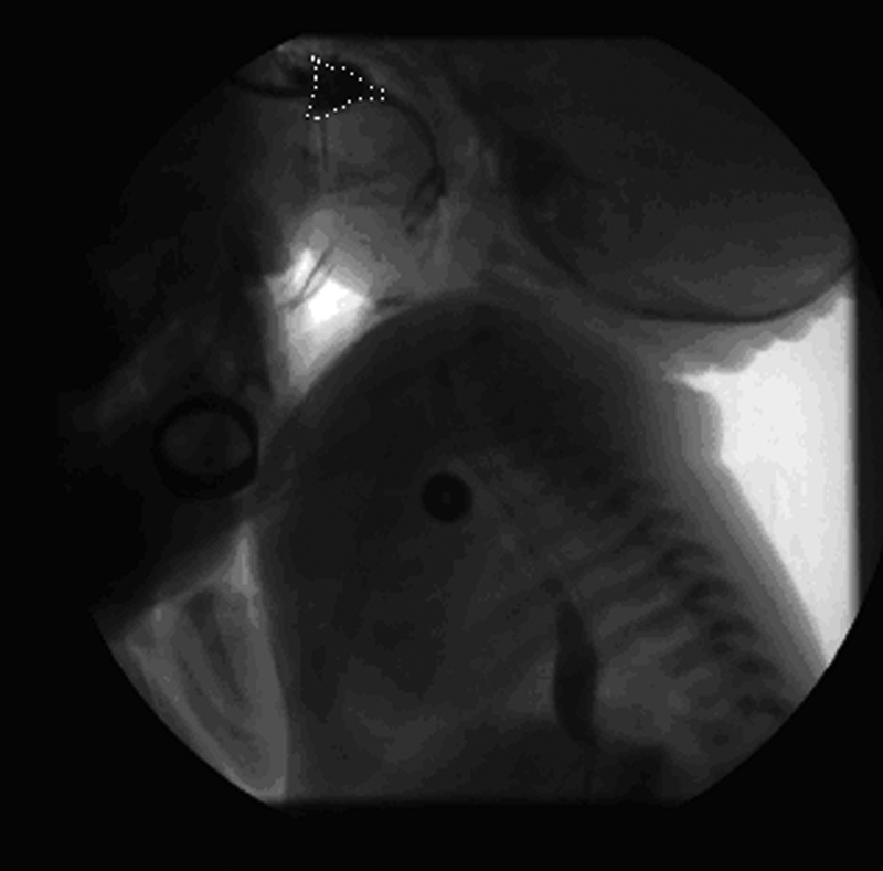
From the data in the present study, which is illustrated in [Figs. 8], [9] and [10], it is possible to verify the presence of esophageal stenosis on breastfeeding, and leakage of liquid to the nasopharynx on bottle-feeding.


The swallowing initiation varied according to the feeding method and in the same subject. There was a prevalence of the trigger occurring in the vallecula for both groups, with a significant difference for the trigger that occurs in the pharyngeal pillars, for bottle-feeding (28%) versus breastfeeding (1%). Previous studies conducted with an adult sample[10] [11] [23] pointed out that the early leakage to the vallecula indicates a poor bolus control. However, in studies with the infant population,[34] it was observed that the pattern of pharyngeal swallowing triggered in the vallecula had a high incidence, and the swallowing could not be triggered at the hypopharynx. This specific finding was not corroborated by the present study. It is difficult to contextualize the variable bolus retention. It was considered as the presence of material collected in the pharyngeal recesses at the same time a respiratory pause occurred, which made it different from bolus retention in a pattern of two, three and even four suctions to trigger a swallow. There was a predominance of bolus retention for the bottle-feeding method (60%), with a smaller incidence for the breastfeeding method (28%). There was a higher occurrence at the end of the feeding for both groups.
There were a few episodes of laryngeal penetration, and most of them happened on the manner of bottle-feeding offer, in 24% of the sample, with clearing in 50% of the times and 4% in the breastfeeding manner. The occurrence of the variable aspiration, differently from other studies that showed high incidence percentages1,[22] [23] [24] presented only 8% in this study with the subjects having, respectively, history of laryngomalacia and prematurity, corrected esophagus atresia and prematurity.
The discrepancy between this finding may be due to differences of the population of present study from the others studies. The sample constitution (all the neonates or infants of the sample were in transition to breastfeeding and were breastfed exclusively) could may suggest minor problems in the oropharyngeal swallowing function.
The description of the enlargement of the hole in the bottle nipple described on previous researches to increase and promote liquid flow may justify the different results from the present study. There is a vast bibliography on the benefits of flow control in the prevention of swallowing problems by reducing the flow, limiting the exit with slow flow holes, and/or thickening the liquid.[35] [36] [37] [38] [39] Given that, it can be concluded that in the translactation technique with urethral catheter number 6 or even 8, the flow is tightly controlled, even with the supposed increase in flow due to the ejection of mother's milk in a normal process of breastfeeding.
Another significant finding that is in line with the findings from other studies[22] [23] [34] is the high incidence of GER related to the symptoms/claims of respiratory problems such as apnea, cyanosis, and respiratory distress not yet explained. The high incidence of GER cannot be solely attributed to the bottle-feeding method (88%), hence, in the present study, the VFSS evaluation always began with breastfeeding, testing the GER at the end of the exam, even though many times the episodes had been observed at different moments. In the present study, all of the variables were compared at the initial and final moments of feeding, with significant differences in bottle-feeding for the nasopharyngeal reflux/velar closure and GER. One can consider the possible impact of fatigue on that. At the end of the evaluation there is a significant gastric fullfilling, thus may explain the greater occurrence of GER at the final moments of the assessment.
As limitations we can consider the fact that this is a retrospective study; therefore, it does not enable stronger control and rigidity with some variables. In the present study, we can point the fact that all subjects were pulled into one group, consisting of term and preterm infants. We can minimize it, considering that the minimum birth age was 32 weeks, with all of them having ≥ 39 weeks of corrected age and 22 days of life at the moment of the evaluation. Remember that each infant was evaluated comparatively in the two feeding ways.
For future studies, we can suggest a randomized method of trial presentation to avoid breast-feeding being always the first method.
It is worth mentioning that investigations with neonates and infants, such as the present study, are commonly performed with a reduced sample size due to the necessary extreme care with the exam indication, trying to avoid radiation exposure.
Conclusion
This study was a comparative analysis of the characteristics of the swallowing function in breastfeeding and bottle-feeding, and significant differences were observed between the two methods, with an impact on the VFSS findings, especially concerning velar function. In the present study, no significant association was observed between the velar disfunction (rhinopharyngeal escape) and the other variables. A total of 88% of the neonates and infants had GER, and this indicates the close link between GER and the symptoms/claims of respiratory problems such as apnea, cyanosis, and respiratory distress.
More studies are necessary for the comprehension of the observed phenomena, enabling the speech and language pathologist to make more accurate diagnoses and develop more accurate approaches to neonatal and infant dysphagia.
No conflict of interest has been declared by the author(s).
-
References
- 1 Bae SO, Lee GP, Seo HG, Oh BM, Han TR. Clinical characteristics associated with aspiration or penetration in children with swallowing problem. Ann Rehabil Med 2014; 38 (06) 734-741
- 2 Vazquez JL, Buonomo C. Feeding difficulties in the first days of life: findings on upper gastrointestinal series and the role of the videofluoroscopic swallowing study. Pediatr Radiol 1999; 29 (12) 894-896
- 3 Pikus L, Levine MS, Yang YX. , et al. Videofluoroscopic studies of swallowing dysfunction and the relative risk of pneumonia. AJR Am J Roentgenol 2003; 180 (06) 1613-1616
- 4 Prasse JE, Kikano GE. An overview of pediatric dysphagia. Clin Pediatr (Phila) 2009; 48 (03) 247-251
- 5 Davis NL, Liu A, Rhein L. Feeding immaturity in preterm neonates: risk factors for oropharyngeal aspiration and timing of maturation. J Pediatr Gastroenterol Nutr 2013; 57 (06) 735-740
- 6 Tutor JD, Srinivasan S, Gosa MM, Spentzas T, Stokes DC. Pulmonary function in infants with swallowing dysfunction. PLoS One 2015; 10 (05) e0123125
- 7 Mello-Filho FV, Mamede RCM, Duca-Silva AP. , et al. Videofluoroscopia da deglutição em crianças. In: Jotz GP, Carrara-De Angelis E, Barros APB. Tratado da deglutição e disfagia: no adulto e na criança. Rio de Janeiro: Revinter; 2009
- 8 Dodrill P, Gosa MM. Pediatric Dysphagia: Physiology, Assessment, and Management. Ann Nutr Metab 2015; 66 (05) (Suppl. 05) 24-31
- 9 Lefton-Greif MA, McGrattan KE, Carson KA, Pinto JM, Wright JM, Martin-Harris B. First Steps Towards Development of an Instrument for the Reproducible Quantification of Oropharyngeal Swallow Physiology in Bottle-Fed Children. Dysphagia 2018; 33 (01) 76-82
- 10 Logeman JA. Evaluation and treatment of swallowing disorders. San Diego (CA): College-Hill Press; 1983
- 11 Arvedson JC. Assessment of pediatric dysphagia and feeding disorders: clinical and instrumental approaches. Dev Disabil Res Rev 2008; 14 (02) 118-127
- 12 American Speech-Language-Hearing Association. Guidelines for Speech-Language Pathologists Performing Videofluoroscopic Swallowing Studies. ASHA Special Interest Division 13, Swallowing and Swallowing Disorders (Dysphagia).2002, 2004
- 13 Martin-Harris B, Jones B. The videofluorographic swallowing study. Phys Med Rehabil Clin N Am 2008; 19 (04) 769-785 , viii
- 14 Weckmueller J, Easterling C, Arvedson J. Preliminary temporal measurement analysis of normal oropharyngeal swallowing in infants and young children. Dysphagia 2011; 26 (02) 135-143
- 15 Goldfield EC, Smith V, Buonomo C, Perez J, Larson K. Preterm infant swallowing of thin and nectar-thick liquids: changes in lingual-palatal coordination and relation to bolus transit. Dysphagia 2013; 28 (02) 234-244 . Doi: 10.1007/s00455-012-9440-y
- 16 Stuart S, Motz JM. Viscosity in infant dysphagia management: comparison of viscosity of thickened liquids used in assessment and thickened liquids used in treatment. Dysphagia 2009; 24 (04) 412-422
- 17 Ekberg O, Stading M, Johansson D, Bülow M, Ekman S, Wendin K. Flow properties of oral contrast medium formulations depend on the temperature. Acta Radiol 2010; 51 (04) 363-367 . Doi: 10.3109/02841851003645751
- 18 López CP, Chiari BM, Goulart AL, Furkim AM, Guedes ZC. Assessment of swallowing in preterm newborns fed by bottle and cup. CoDAS 2014; 26 (01) 81-86
- 19 Cichero J, Nicholson T, Dodrill P. Liquid barium is not representative of infant formula: characterisation of rheological and material properties. Dysphagia 2011; 26 (03) 264-271
- 20 Frazier J, Chestnut AH, Jackson A, Barbon CE, Steele CM, Pickler L. Pickler l. Understanding the viscosity of liquids used in infant dysphagia management. Dysphagia 2016; 31 (05) 672-679
- 21 Lima GM S. Métodos especiais de alimentação: copinho – relactação – translactação. In: REGO, J. D. Aleitamento materno. São Paulo, Rio de Janeiro, Belo Horizonte: Atheneu; 2000: 265-278
- 22 Elad D, Kozlovsky P, Blum O. , et al. Biomechanics of milk extraction during breast-feeding. Proc Natl Acad Sci U S A 2014; 111 (14) 5230-5235
- 23 Mercado-Deane MG, Burton EM, Harlow SA. , et al. Swallowing dysfunction in infants less than 1 year of age. Pediatr Radiol 2001; 31 (06) 423-428
- 24 Hayoun P, Engmann J, Mowlavi S, Le Reverend B, Burbidge A, Ramaioli M. A model experiment to understand the oral phase of swallowing of Newtonian liquids. J Biomech 2015; 48 (14) 3922-3928
- 25 Schrank W, Al-Sayed LE, Beahm PH, Thach BT. Feeding responses to free-flow formula in term and preterm infants. J Pediatr 1998; 132 (3 Pt 1): 426-430
- 26 German RZ, Crompton AW, Owerkowicz T, Thexton AJ. Volume and rate of milk delivery as determinants of swallowing in an infant model animal (Sus scrofia). Dysphagia 2004; 19 (03) 147-154
- 27 Ardran GM, Kemp FH, Lind J. A Cineradiographic study of breast feeding. Br J Radiol 1958; 31 (363) 156-162
- 28 Weber F, Woolridge MW, Baum JD. An ultrasonographic study of the organisation of sucking and swallowing by newborn infants. Dev Med Child Neurol 1986; 28 (01) 19-24
- 29 Bosma JF, Hepburn LG, Josell SD, Baker K. Ultrasound demonstration of tongue motions during suckle feeding. Dev Med Child Neurol 1990; 32 (03) 223-229
- 30 Eishima K. The analysis of sucking behaviour in newborn infants. Early Hum Dev 1991; 27 (03) 163-173
- 31 Geddes DT, Sakalidis VS. Ultrasound Imaging of Breastfeeding--A Window to the Inside: Methodology, Normal Appearances, and Application. J Hum Lact 2016; 32 (02) 340-349 . Doi: 10.1177/0890334415626152 Review
- 32 Sakalidis VS, Geddes DT. Suck-Swallow-Breathe Dynamics in Breastfed Infants. J Hum Lact 2016; 32 (02) 201-211 , quiz 393–395
- 33 Nowak AJ, Smith WL, Erenberg A. Imaging evaluation of artificial nipples during bottle feeding. Arch Pediatr Adolesc Med 1994; 148 (01) 40-42
- 34 Newman LA, Keckley C, Petersen MC, Hamner A. Swallowing function and medical diagnoses in infants suspected of Dysphagia. Pediatrics 2001; 108 (06) E106
- 35 Goldfield EC, Buonomo C, Fletcher K. , et al. Premature infant swallowing: patterns of tongue-soft palate coordination based upon videofluoroscopy. Infant Behav Dev 2010; 33 (02) 209-218
- 36 Cassiani RA, Santos CM, Parreira LC, Dantas RO. The relationship between the oral and pharyngeal phases of swallowing. Clinics (São Paulo) 2011; 66 (08) 1385-1388
- 37 Rossi MS, Buhler KE, Ventura GA, Otoch JP, Limongi SC. Laryngeal cleft type I in neonate: case report. CoDAS 2014; 26 (05) 421-424
- 38 Leonard RJ, White C, McKenzie S, Belafsky PC. Effects of bolus rheology on aspiration in patients with Dysphagia. J Acad Nutr Diet 2014; 114 (04) 590-594
- 39 Newman R, Vilardell N, Clavé P, Speyer R. Effect of Bolus Viscosity on the Safety and Efficacy of Swallowing and the Kinematics of the Swallow Response in Patients with Oropharyngeal Dysphagia: White Paper by the European Society for Swallowing Disorders (ESSD). Dysphagia 2016; 31 (02) 232-249 . Doi: 10.1007/s00455-016-9696-8
Address for correspondence
-
References
- 1 Bae SO, Lee GP, Seo HG, Oh BM, Han TR. Clinical characteristics associated with aspiration or penetration in children with swallowing problem. Ann Rehabil Med 2014; 38 (06) 734-741
- 2 Vazquez JL, Buonomo C. Feeding difficulties in the first days of life: findings on upper gastrointestinal series and the role of the videofluoroscopic swallowing study. Pediatr Radiol 1999; 29 (12) 894-896
- 3 Pikus L, Levine MS, Yang YX. , et al. Videofluoroscopic studies of swallowing dysfunction and the relative risk of pneumonia. AJR Am J Roentgenol 2003; 180 (06) 1613-1616
- 4 Prasse JE, Kikano GE. An overview of pediatric dysphagia. Clin Pediatr (Phila) 2009; 48 (03) 247-251
- 5 Davis NL, Liu A, Rhein L. Feeding immaturity in preterm neonates: risk factors for oropharyngeal aspiration and timing of maturation. J Pediatr Gastroenterol Nutr 2013; 57 (06) 735-740
- 6 Tutor JD, Srinivasan S, Gosa MM, Spentzas T, Stokes DC. Pulmonary function in infants with swallowing dysfunction. PLoS One 2015; 10 (05) e0123125
- 7 Mello-Filho FV, Mamede RCM, Duca-Silva AP. , et al. Videofluoroscopia da deglutição em crianças. In: Jotz GP, Carrara-De Angelis E, Barros APB. Tratado da deglutição e disfagia: no adulto e na criança. Rio de Janeiro: Revinter; 2009
- 8 Dodrill P, Gosa MM. Pediatric Dysphagia: Physiology, Assessment, and Management. Ann Nutr Metab 2015; 66 (05) (Suppl. 05) 24-31
- 9 Lefton-Greif MA, McGrattan KE, Carson KA, Pinto JM, Wright JM, Martin-Harris B. First Steps Towards Development of an Instrument for the Reproducible Quantification of Oropharyngeal Swallow Physiology in Bottle-Fed Children. Dysphagia 2018; 33 (01) 76-82
- 10 Logeman JA. Evaluation and treatment of swallowing disorders. San Diego (CA): College-Hill Press; 1983
- 11 Arvedson JC. Assessment of pediatric dysphagia and feeding disorders: clinical and instrumental approaches. Dev Disabil Res Rev 2008; 14 (02) 118-127
- 12 American Speech-Language-Hearing Association. Guidelines for Speech-Language Pathologists Performing Videofluoroscopic Swallowing Studies. ASHA Special Interest Division 13, Swallowing and Swallowing Disorders (Dysphagia).2002, 2004
- 13 Martin-Harris B, Jones B. The videofluorographic swallowing study. Phys Med Rehabil Clin N Am 2008; 19 (04) 769-785 , viii
- 14 Weckmueller J, Easterling C, Arvedson J. Preliminary temporal measurement analysis of normal oropharyngeal swallowing in infants and young children. Dysphagia 2011; 26 (02) 135-143
- 15 Goldfield EC, Smith V, Buonomo C, Perez J, Larson K. Preterm infant swallowing of thin and nectar-thick liquids: changes in lingual-palatal coordination and relation to bolus transit. Dysphagia 2013; 28 (02) 234-244 . Doi: 10.1007/s00455-012-9440-y
- 16 Stuart S, Motz JM. Viscosity in infant dysphagia management: comparison of viscosity of thickened liquids used in assessment and thickened liquids used in treatment. Dysphagia 2009; 24 (04) 412-422
- 17 Ekberg O, Stading M, Johansson D, Bülow M, Ekman S, Wendin K. Flow properties of oral contrast medium formulations depend on the temperature. Acta Radiol 2010; 51 (04) 363-367 . Doi: 10.3109/02841851003645751
- 18 López CP, Chiari BM, Goulart AL, Furkim AM, Guedes ZC. Assessment of swallowing in preterm newborns fed by bottle and cup. CoDAS 2014; 26 (01) 81-86
- 19 Cichero J, Nicholson T, Dodrill P. Liquid barium is not representative of infant formula: characterisation of rheological and material properties. Dysphagia 2011; 26 (03) 264-271
- 20 Frazier J, Chestnut AH, Jackson A, Barbon CE, Steele CM, Pickler L. Pickler l. Understanding the viscosity of liquids used in infant dysphagia management. Dysphagia 2016; 31 (05) 672-679
- 21 Lima GM S. Métodos especiais de alimentação: copinho – relactação – translactação. In: REGO, J. D. Aleitamento materno. São Paulo, Rio de Janeiro, Belo Horizonte: Atheneu; 2000: 265-278
- 22 Elad D, Kozlovsky P, Blum O. , et al. Biomechanics of milk extraction during breast-feeding. Proc Natl Acad Sci U S A 2014; 111 (14) 5230-5235
- 23 Mercado-Deane MG, Burton EM, Harlow SA. , et al. Swallowing dysfunction in infants less than 1 year of age. Pediatr Radiol 2001; 31 (06) 423-428
- 24 Hayoun P, Engmann J, Mowlavi S, Le Reverend B, Burbidge A, Ramaioli M. A model experiment to understand the oral phase of swallowing of Newtonian liquids. J Biomech 2015; 48 (14) 3922-3928
- 25 Schrank W, Al-Sayed LE, Beahm PH, Thach BT. Feeding responses to free-flow formula in term and preterm infants. J Pediatr 1998; 132 (3 Pt 1): 426-430
- 26 German RZ, Crompton AW, Owerkowicz T, Thexton AJ. Volume and rate of milk delivery as determinants of swallowing in an infant model animal (Sus scrofia). Dysphagia 2004; 19 (03) 147-154
- 27 Ardran GM, Kemp FH, Lind J. A Cineradiographic study of breast feeding. Br J Radiol 1958; 31 (363) 156-162
- 28 Weber F, Woolridge MW, Baum JD. An ultrasonographic study of the organisation of sucking and swallowing by newborn infants. Dev Med Child Neurol 1986; 28 (01) 19-24
- 29 Bosma JF, Hepburn LG, Josell SD, Baker K. Ultrasound demonstration of tongue motions during suckle feeding. Dev Med Child Neurol 1990; 32 (03) 223-229
- 30 Eishima K. The analysis of sucking behaviour in newborn infants. Early Hum Dev 1991; 27 (03) 163-173
- 31 Geddes DT, Sakalidis VS. Ultrasound Imaging of Breastfeeding--A Window to the Inside: Methodology, Normal Appearances, and Application. J Hum Lact 2016; 32 (02) 340-349 . Doi: 10.1177/0890334415626152 Review
- 32 Sakalidis VS, Geddes DT. Suck-Swallow-Breathe Dynamics in Breastfed Infants. J Hum Lact 2016; 32 (02) 201-211 , quiz 393–395
- 33 Nowak AJ, Smith WL, Erenberg A. Imaging evaluation of artificial nipples during bottle feeding. Arch Pediatr Adolesc Med 1994; 148 (01) 40-42
- 34 Newman LA, Keckley C, Petersen MC, Hamner A. Swallowing function and medical diagnoses in infants suspected of Dysphagia. Pediatrics 2001; 108 (06) E106
- 35 Goldfield EC, Buonomo C, Fletcher K. , et al. Premature infant swallowing: patterns of tongue-soft palate coordination based upon videofluoroscopy. Infant Behav Dev 2010; 33 (02) 209-218
- 36 Cassiani RA, Santos CM, Parreira LC, Dantas RO. The relationship between the oral and pharyngeal phases of swallowing. Clinics (São Paulo) 2011; 66 (08) 1385-1388
- 37 Rossi MS, Buhler KE, Ventura GA, Otoch JP, Limongi SC. Laryngeal cleft type I in neonate: case report. CoDAS 2014; 26 (05) 421-424
- 38 Leonard RJ, White C, McKenzie S, Belafsky PC. Effects of bolus rheology on aspiration in patients with Dysphagia. J Acad Nutr Diet 2014; 114 (04) 590-594
- 39 Newman R, Vilardell N, Clavé P, Speyer R. Effect of Bolus Viscosity on the Safety and Efficacy of Swallowing and the Kinematics of the Swallow Response in Patients with Oropharyngeal Dysphagia: White Paper by the European Society for Swallowing Disorders (ESSD). Dysphagia 2016; 31 (02) 232-249 . Doi: 10.1007/s00455-016-9696-8